Sample information |
|
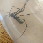
| Picture |
|
|---|---|
| Location | |
| Collection date | 08/15/2025 |
| Captive / Cultivated? | Wild-caught |
| Group | Edmund Burke School |
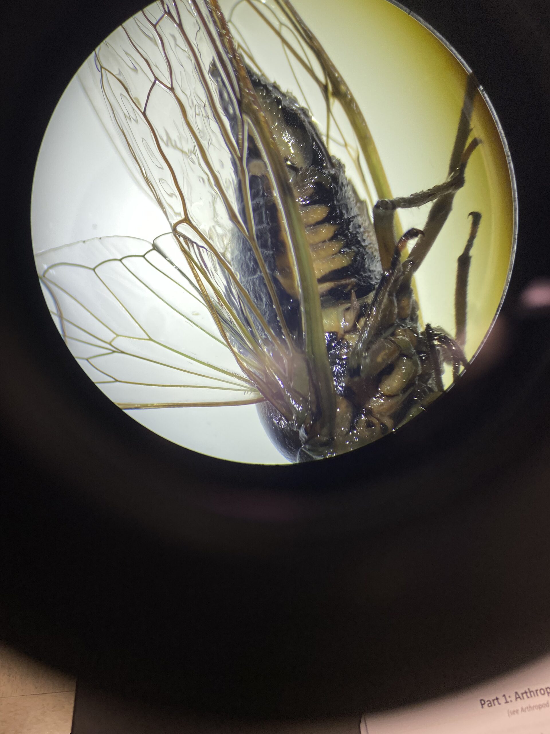
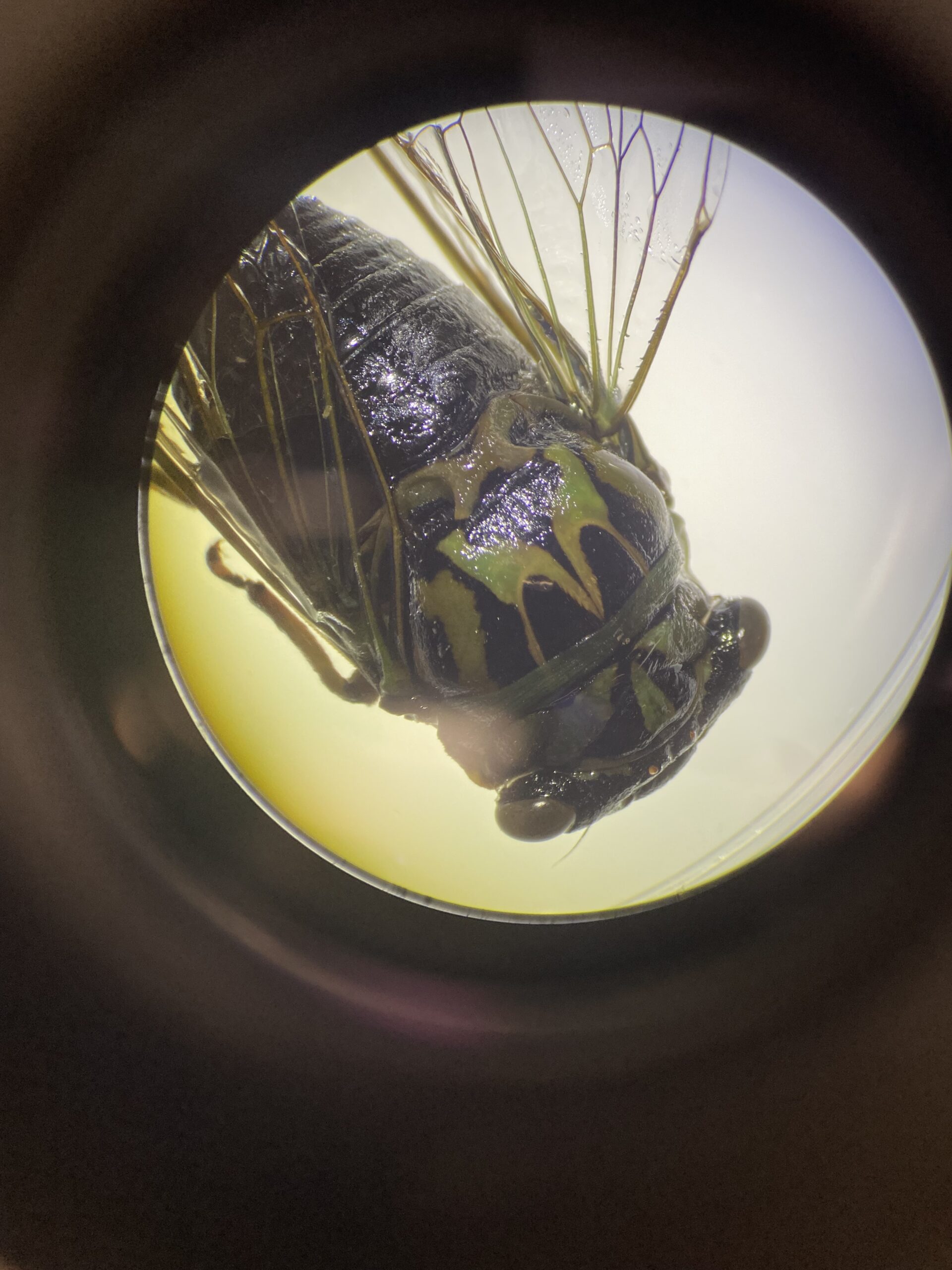
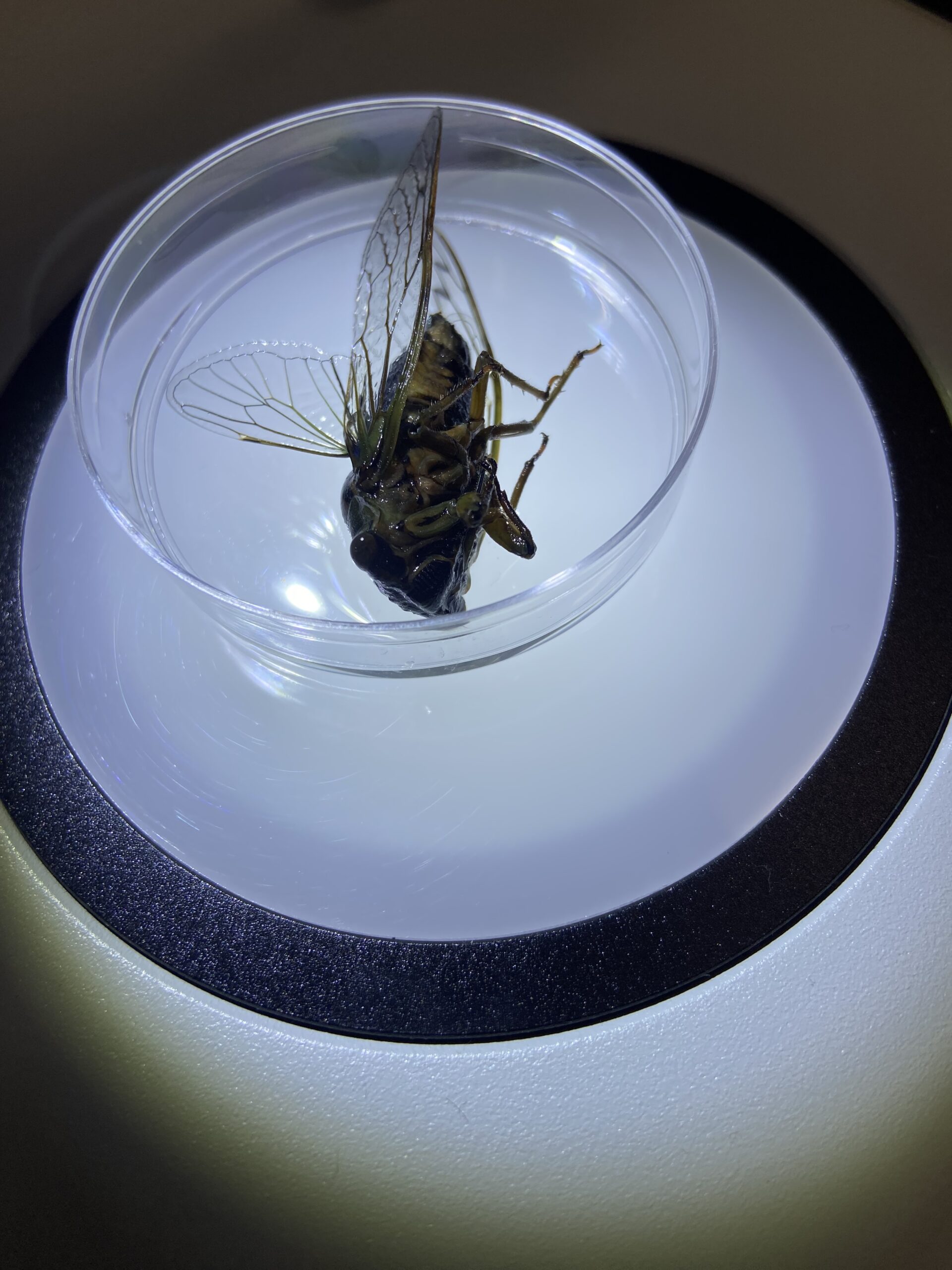
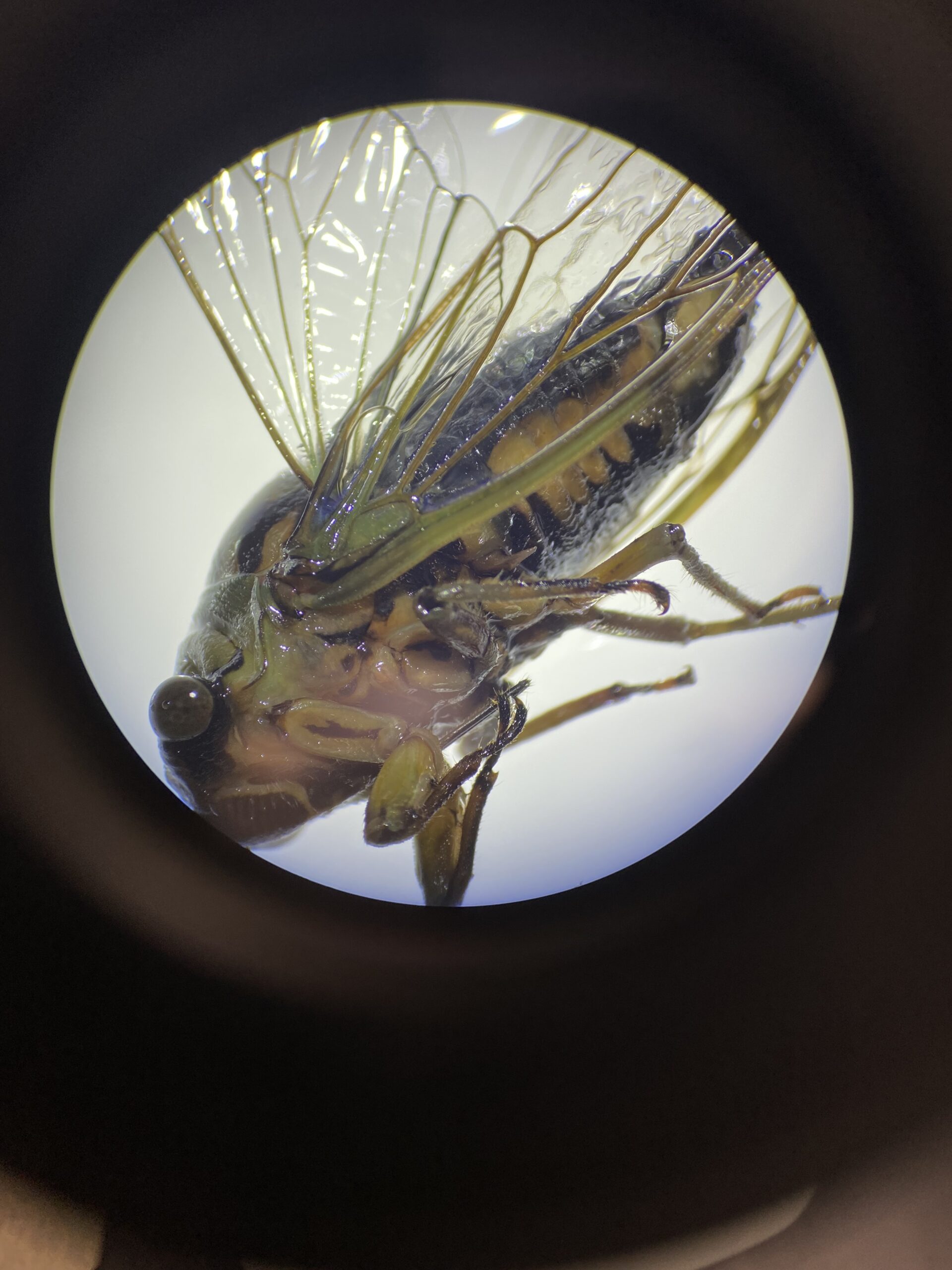
| Observations | Caught outside on a driveway in a fairly wooded, suburban area. Caught in late summer. Weather was cloudy, 89 degrees, with 63% humidity. It seemed old and was less squirmy than the other cicada. In comparison, its colors were more muted and less shiny. Top wing on each side is tall and somewhat narrow, standing straight up from the side of the thorax. Bottom wing is shorter and longer, sitting against the side of the abdomen. All wings are very thin. Wings are translucent with strong green and yellow veins. Some edges of the wings are veined, with an especially thick vein on the bottom edge of the bottom wing. It has 4 wings and 6 legs. The foreleg has a thick ovular upper part and a much thinner lower part of the leg. The lower part of the foreleg is bent towards the abdomen. The hind legs has a thicker cylindrical upper part of the leg and thinner lower part that has small hairs. Hind legs appear longer than forelegs. The head is flat with a rounded, triangular face. The mouth is in the center of the face and takes up most of it. It is ridged and roughly rectangular. It has two eyes that are large and spherical. They are facing forward on either side of the face and protrude significantly. It has no antennae. The body has three regions and is roughly cylindrical with a smaller head. The abdomen is thickest closest to the thorax and the tapers off significantly. The top is mostly black with patches of green on the thorax and head. The bottom is brown and black. The legs and wing veins are green and brown. |
| Putative identification | Arthropoda Insecta Hemiptera |
Methods |
|
| Extraction kit | DNeasy (Qiagen) |
| DNA extraction location | Abdomen |
| Single or Duplex PCR | Single Reaction |
| Gel electrophoresis system | MiniOne |
| Buffer | TBE |
| DNA stain | GelGreen |
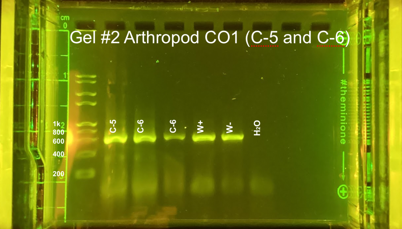
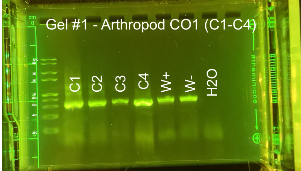
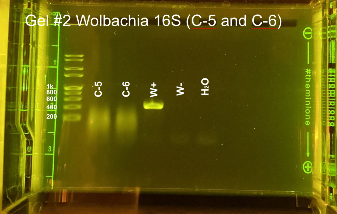
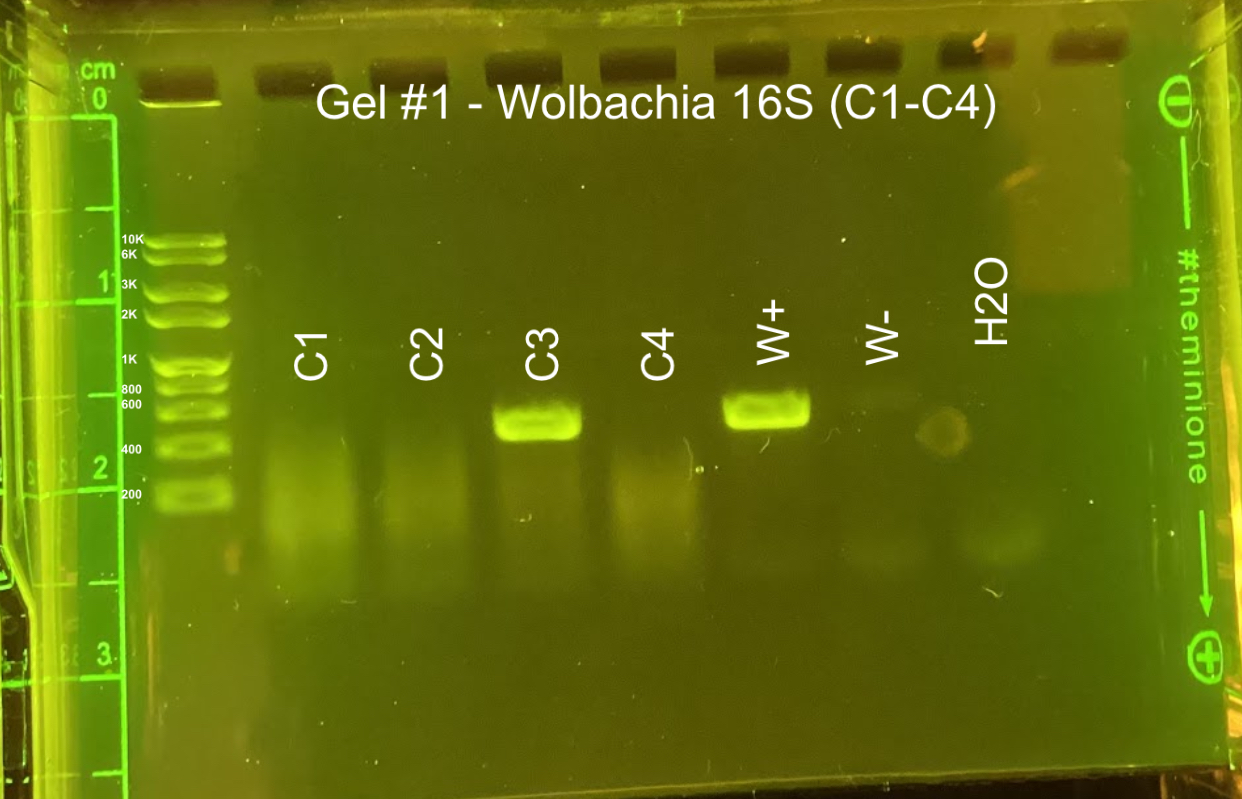
| Gel images |
|
| Protocol notes | DNA Extraction: went as planned PCR: went as planned Gel electrophoresis, Arthropod CO1: gel #1 lanes – ladder, C-1, C-2, C-3, C-4, positive Wolbachia control, negative Wolbachia control, water control; gel #2 lanes – ladder, C-5, C-6, C-6 again, Wolbachia positive control, Wolbachia negative control, water control We accidentally added C-6 DNA to two wells. All of the samples and the arthropod controls had DNA, but the water control did not. Gel electrophoresis, Wolbachia 16S: gel #1 lanes – ladder, C-1, C-2, C-3, C-4, positive Wolbachia control, negative Wolbachia control, water control; gel #2 lanes – ladder, C-5, C-6 again, Wolbachia positive control, Wolbachia negative control, water control We only added 3 microliters of W- on gel 2. Both W+ had Wolbachia DNA as did C-3. DNA sequencing: Sequencing did not work. We are assuming it its genus is Neotibicin based on morphology. |
Results |
|
| Wolbachia presence | No |
| Confidence level | High |
| Explanation of confidence level | All of the samples had a band for arthropod CO1 DNA. The Wolbachia positive control had a band for both arthropod and Wolbachia DNA. The Wolbachia negative control only had a band for arthropod DNA. The water control did not have DNA in any gel. This shows that we did not have any contamination. |
| Wolbachia 16S sequence | |
| Arthropod COI sequence |
|
| Summary | The Hemiptera was found to be negative for Wolbachia. |
 Joro Spider_AG wolbochia_hendricks_lab
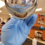 Millipede_AG wolbochia_lab_hendricks
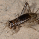 Japanese Burrowing Cricket, State College, PA – Wolbachia Results
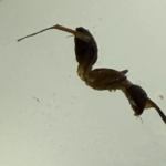 Ant like creature – Draft
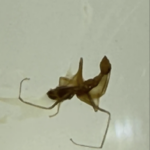 Myrmaplata plantaleoides (Unsure) – Draft