| Protocol notes |
PCR notes:
Purpose: To extend the DNA samples that we retrieved by increasing the amount of DNA by producing copies of the DNA.
Materials:
- Thermal cycler
- Vortex
- Mini-centrifuge
- Gloves
- Squirt bottle
- 0.2 mL PCR tubes
- Rack for 0.2 mL PCR tubes
- 1.5 mL tubes
- Rack for 1.5 mL
- Balance tube
- Disposal cup for tips
- Pipettes (200µL and 20µL)
- Sharpie
- DNA samples from arthropod specimens
- +/- Wolbachia controls
- +DNA control
- Sterile, nuclease-free water
- Taq Master Mix
- Wolbachia_F and Wolbachia_R primers
Methods:
- A teacher had prepared the Thermal cycler and PCR program was entered
- All unnecessary items were removed from lab
- Nitrile gloves were put on and all surfaces were wiped down with 70% Ethanol.
- 1.5 mL microcentrifuge tubes were collected. Tube was labeled with “PCR” tube was placed on a 1.5mL tube rack
- Eight 0.2 mL PCR tubes were collected. Tubes were labeled and numbered with initials. Tubes placed in PCR tube rack.
- P-20 pipette were used and 2µL of template DNA were added to corresponding PCR tube. Tips were changed between each tube.
- PCR tubes were set aside. Templated DNA tubes were placed into storage
- P-200 pipette were used and reagents from the 9 reactions were added to the 1.5mL tube marked “PCR”. Tips were changed between each reagent and reagent were checked off after it was added.
- Tube was closed and briefly vortexed for 5 seconds
- PCR tube was placed on one side of the mini-centrifuge and Balancer tube on opposite sides. PCR Cocktail was quickly spun down for 4 seconds.
- P-200 pipette was used and 23 µL of PCR was added to tubes 1-8. Tips were changed between each tube.
- Lids were secured on each tube
- PCR tube rotor was used on mini-centrifuge. Tubes 1-4 were centered on one side and 5-8 on the other. Liquids were briefly spun to collect liquid
- Tubes were transferred to thermal cycler.
- Lab station was cleaned and surfaces were wiped with ethanol.
Gel Electrophoresis:
Purpose: To create a agarose gel that allows us to determine whether or not our samples contain Wolbachia. By
analyzing the distance the DNA strand traveled.
Materials:
- Electrophoresis tank
- Power supply
- Standard Electrophoresis casting tray and comb
- Microwave
- Imager
- Electronic balance
- Squirt bottle
- Gloves
- Oven mitt
- Eye protection
- Weigh Dish
- Running Buffer TAE
- 250mL Erlenmeyer flask
- Graduated cylinder
- Rack
- 0.2mL PCR tubes
- 20 microliter pipette
- 20 microliter pipette tips
- Waste cup
- Sharpie
- Agarose powder
- DNA stain
- PCR products
- DNA ladder
Methods:
- As class, 1 working solution of electrophoresis running buffer (TAE) was prepared
- All unnecessary equipment was removed from lab.
- Nitrile gloves were put on and all surfaces were cleaned with 70% ethanol
Prepare the Gel with DNA Stain:
- 0.5g were measured and added to 250 mL flask
- 50 mL of running buffer were added to 250mL flask
- Parafilm was initially used for the microwaving (film melted after 60 seconds)
- Material was switched to Kimwipe for a 30 second interval excess film was cleaned off. The agarose was melted into Buffer/Solvent
- The solution was cooled to about 50-55C, flask was swirled occasion until it was warm to touch.
- Casting tray ends were sealed by placing it in to the gel electrophoresis tank
- 10 microliters were added to agarose. Was swirled to mix
- Comb was selected that accommodated all samples. Combs were placed in gel casting tray
- The melted agarose was slowly poured into the casting tray.
- Gel was allowed to cool undisturbed on a solid, flat surface for 20 minute s until it was solid
- Combs were carefully pulled out and the casting tray was lifted out of the electrophoresis chamber.
- Gel was placed in the electrophoresis chamber with well oriented near the negative electrode.
- Running buffer was estimated and added over the gel.
Prepare to load the Gel:
- Loading Key was fill out on the next page
- 2µL drops of 5X loading buffer were pipetted onto a piece of Parafilm. 10µL were added from each PCR reaction of loading buffer. Mixed well by pipetting up and down several times until the color of the liquid is homogenous.
- 10µL of the DNA ladder was pipetted into the first well. Pipette was hovered above well, pipette was slowly emptied.
- Pipetted carefully 10µL of each PCR sample in different wells. Tips were changed between each sample.
Run the Gel:
- Lid was placed on the gel box, electrodes were connected appropriately.
- Power supply was turned to 125 volts.
- Current was checked to make sure it ran through buffer
- Current was checked to be moving in the correct direction.
- Power ran until the yellow was 3/4 down the gel. Power was then turned off and electrodes were disconnected
lid was removed along with gel using gloves
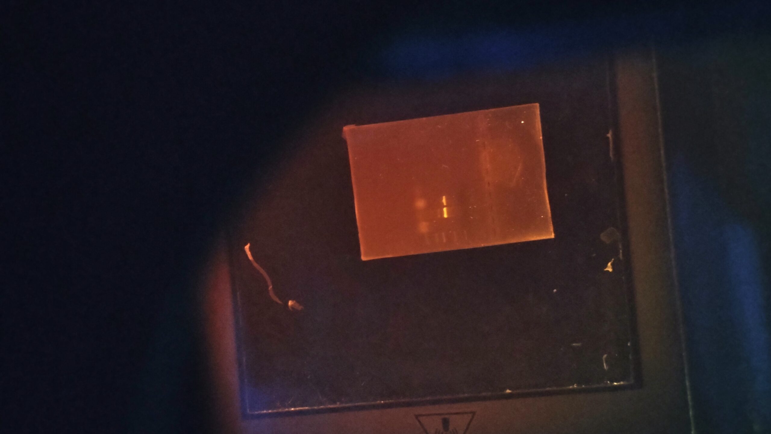
Obtain an image of the Gel:
- Gel was placed on the transilluminator. Eye protection was put on.
- Prescence and absence of bands were noted in each lane.
- Results were documented in lab notebook and a picture of gel was taken
- Used tips were discarded and bench was wiped down.
- Gel images were transferred to computer.
|
| Explanation of confidence level |
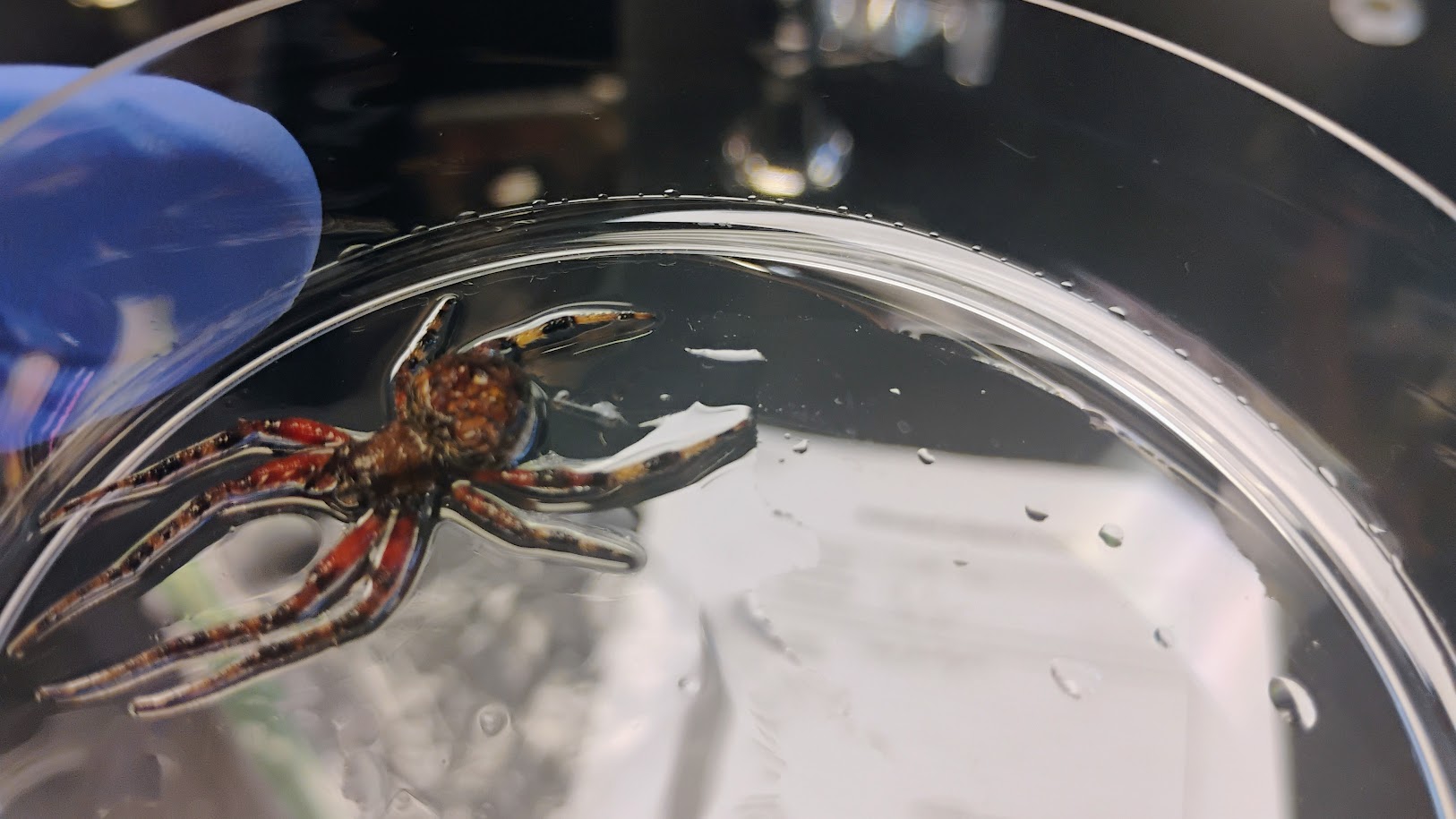
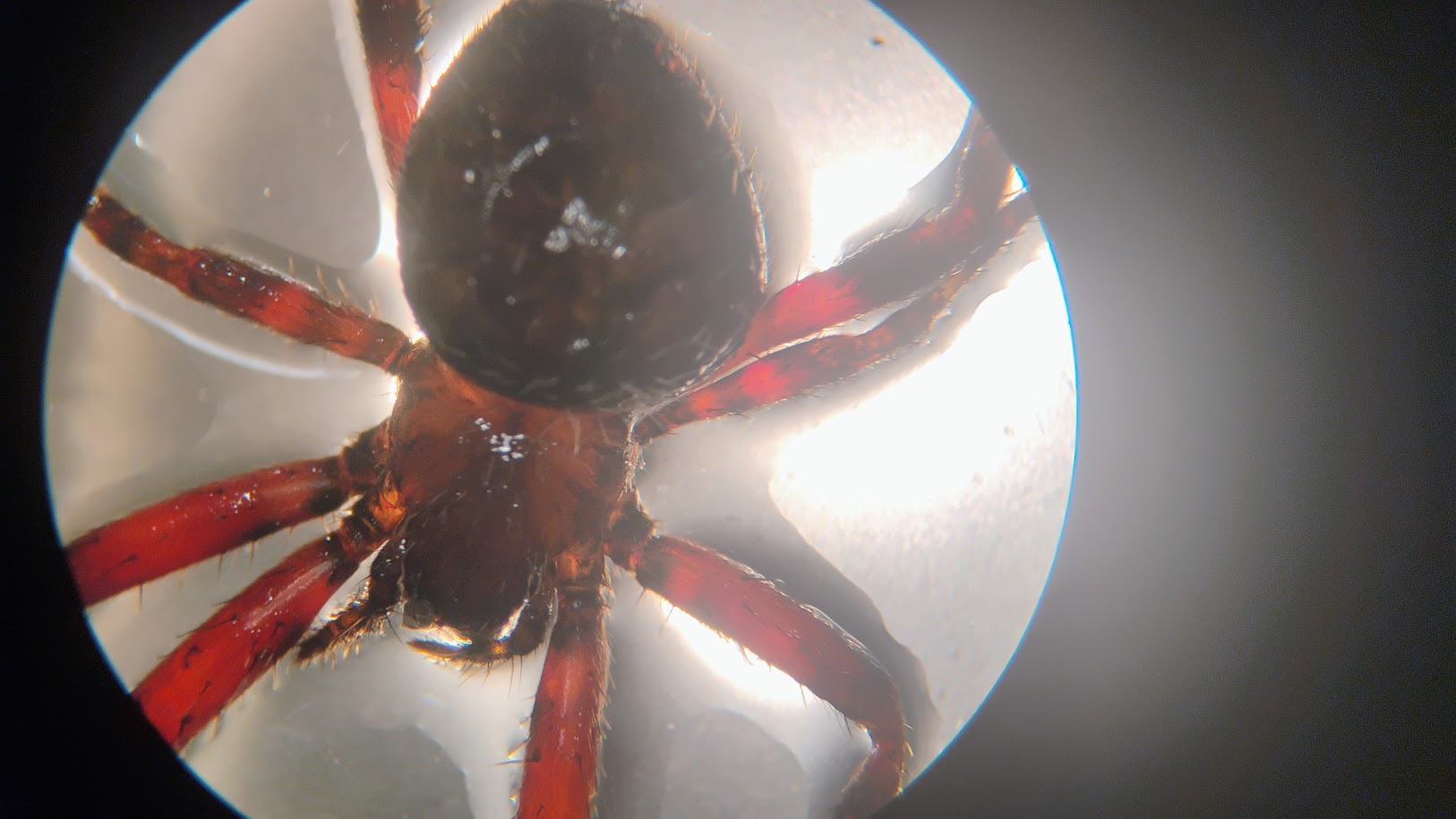
While it is possible that the Red-legged Orb weaver tested positive, it was mixed within a batch of differing arthropods. Unfortunately, before the Gel Electrophoresis, the samples weren’t labeled, causing there to be uncertainty within the experiment itself, and our results to be unknown. All that we do know is that each of the controls did indeed work, but there is obscurity in the results of the additional arthropoda.
|
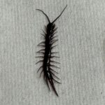 Centipede – MJAR
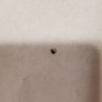 Fruitfly – MJAR
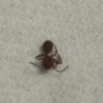 Ant – MJAR
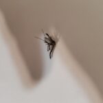 Mosquito – MJAR
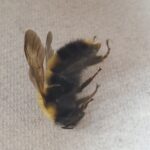 Bumblebee – MJAR