Sample information |
|
| Picture |
|
|---|---|
| Location | |
| Collection date | 05/13/2025 |
| Captive / Cultivated? | Wild-caught |
| Group | California Academy of Mathematics and Science |
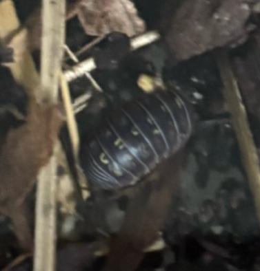
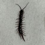
| Observations | This arthropod was collected from a pot in a garden. Black colored exoskeleton with yellow designs in the center of its body. Segmented body, about 1.75 cm long. |
| Putative identification | Arthropoda Malacostraca Isopoda Armadillidae Armadillidium Armadillidium vulgare |
Methods |
|
| Extraction kit | DNeasy (Qiagen) |
| DNA extraction location | Abdomen |
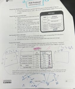
| Single or Duplex PCR | Duplex Reaction |
| Gel electrophoresis system | Standard electrophoresis system |
| Buffer | 1X TAE |
| DNA stain | 100X Fast Blast |
| Gel images |
|
| Protocol notes |
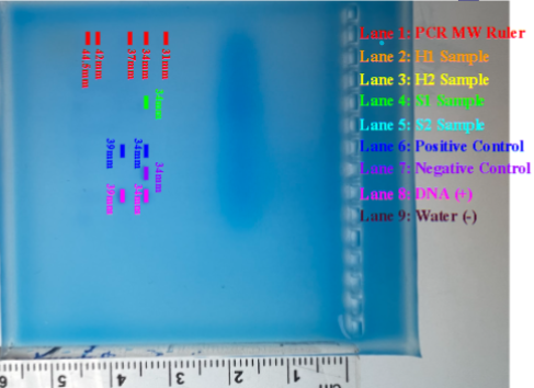
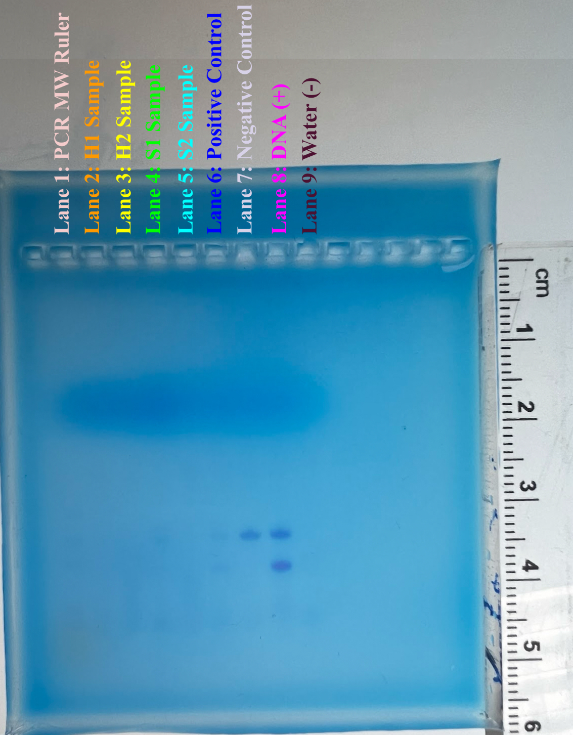
All lanes not labeled do not contain anything in them. This specimen’s bands can be located on the labeled lane 3. |
Results |
|
| Wolbachia presence | Unknown |
| Confidence level | Low |
| Explanation of confidence level | Due to a possible error when performing DNA extraction, no results were visible in our gel after electrophoresis, except for the provided controls. Thus, an unsuccessful lab was performed. Out of the four Armadillidium Vulgare samples, only one had a band indicating presence of Arthropod CO1 gene, and none had any bands indicating presence of Wolbachia 16S rRNA gene. |
| Wolbachia 16S sequence | |
| Arthropod COI sequence |
|
| Summary | |

 Centipede – MJAR
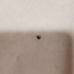 Fruitfly – MJAR
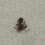 Ant – MJAR
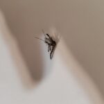 Mosquito – MJAR
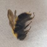 Bumblebee – MJAR