Sample information |
|
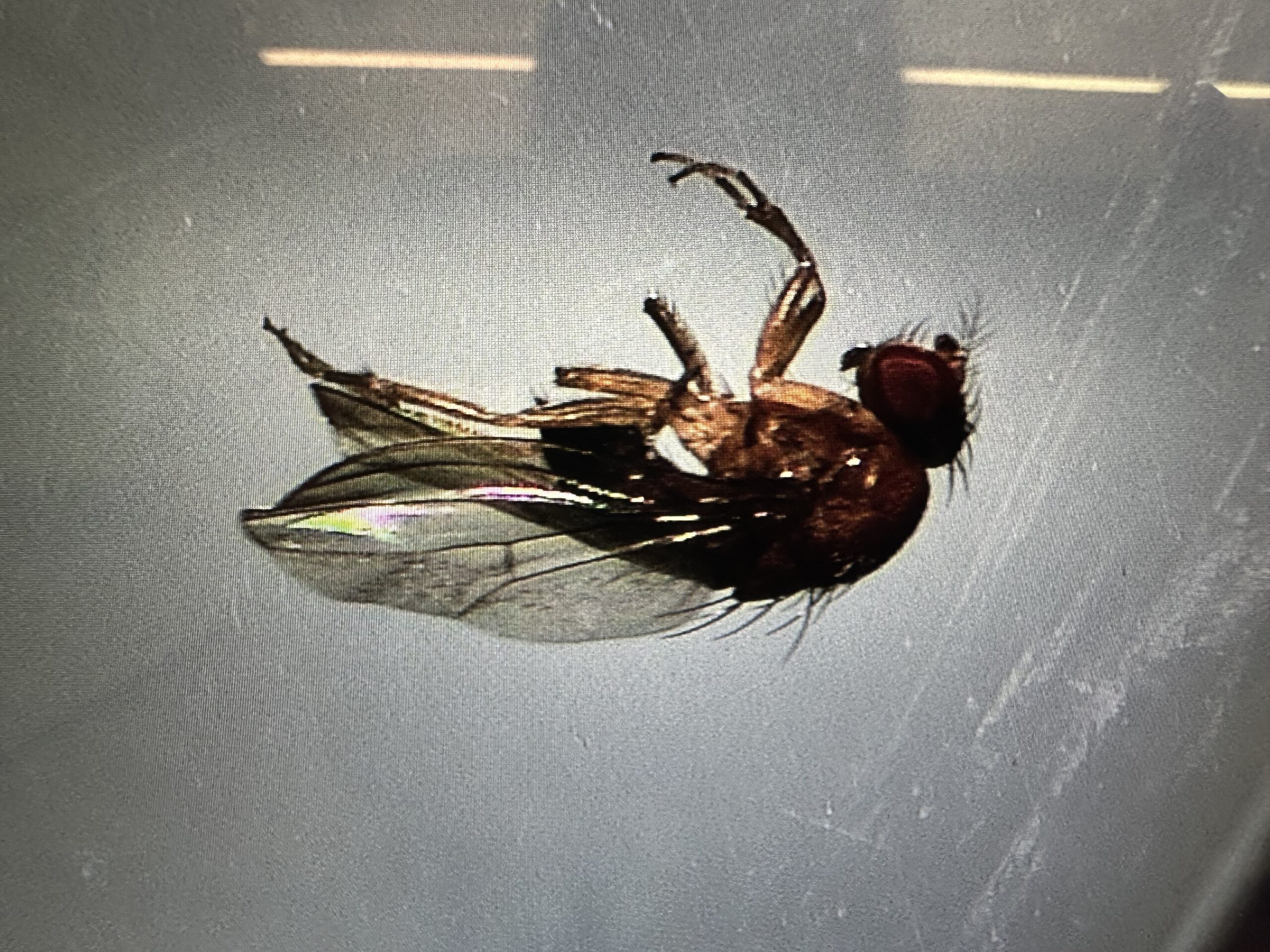
| Picture |
|
|---|---|
| Location | |
| Collection date | 10/02/2025 |
| Captive / Cultivated? | Wild-caught |
| Group | Berkshire Community College |
| Observations | Residential backyard, near some blueberry bushes and a tree stump. By a garden with some rotting tomatoes on the vine. |
| Putative identification | Arthropoda Insecta Diptera Drosophilidae Drosophila Drosophila suzukii |
Methods |
|
| Extraction kit | Monarch DNA extraction (NEB) |
| DNA extraction location | Whole arthropod |
| Single or Duplex PCR | Duplex Reaction |
| Gel electrophoresis system | Edvotek Gel Electrophoresis |
| Buffer | 1X TAE |
| DNA stain | SYBR Safe |
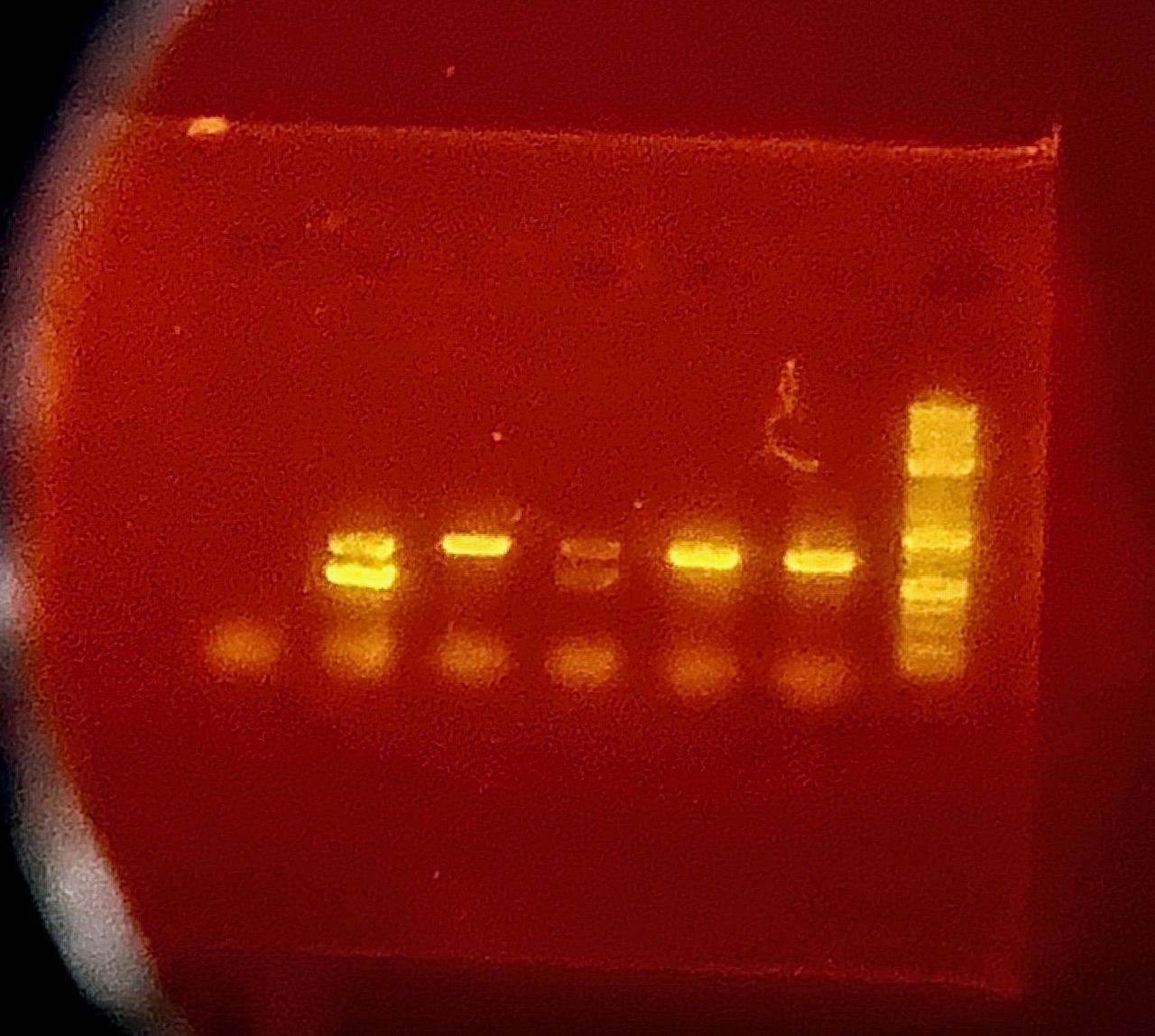
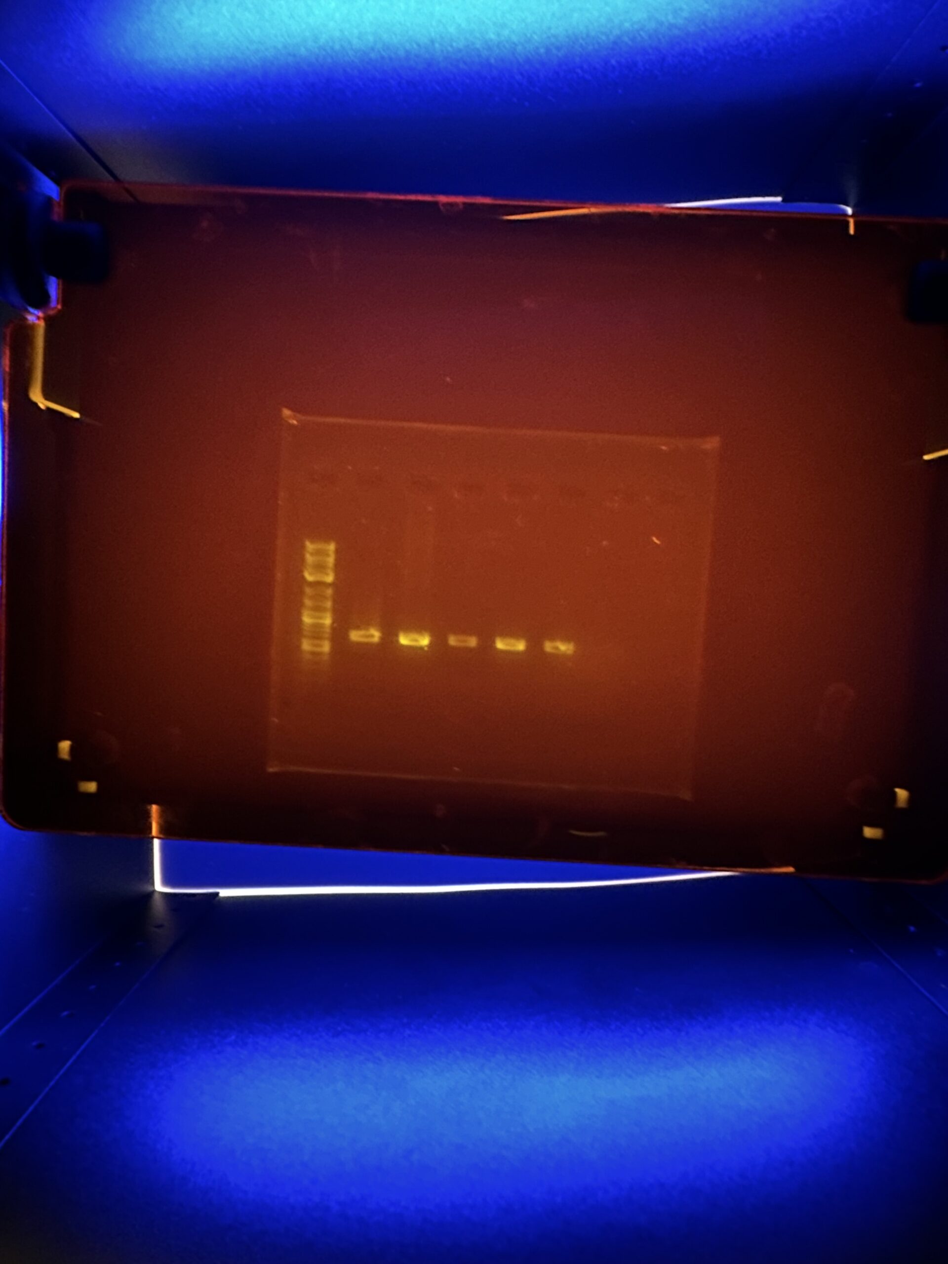
| Gel images |

|
| Protocol notes | We used a DNA extraction protocol based on the insect adaptation of New England’s biolabs’ Monarch Spin gDNA Extraction kit. the specimen was incubated for 30 minutes in a hot water back at 56 degrees C. Our first Gel image was taken on 10/23/2025. It was run at 125 volts for 29 minutes. The DNA Ladder used was New England Biolabs 1kb Plus DNA Ladder for Safe Stains (product#N0559S We ran a second PCR – this time a single PCR and not a duplex – on 11/5/2025. A second gel electrophoresis was run on 11/6/2025. It was run at 125 volts for 25 minutes. The DNA Ladder used was New England Biolabs 1kb Plus DNA Ladder for Safe Stains (product#N0559S). |
Results |
|
| Wolbachia presence | No |
| Confidence level | High |
| Explanation of confidence level | My confidence level is high that my arthropod was tested negative for Wolbachia because all other control samples were clear and accurate. The color and brightness for my and my partners arthropod to have the Arthropod DNA line visible but not Wolbachia line was very clear and obvious. The second gel was ran from a PCR with Arthropod primer and not Wolbachia primer. The intention was to try a different method since my confidence was very high no Wolbachia was present. |
| Wolbachia 16S sequence | |
| Arthropod COI sequence | Download AB1
GAGCTTGAGCCGGAATAGTGGGAACATCTCTAAGAATTTTAATTCGAGCTGAATTAGGTCATCCAGGAGCATTAATTGGAGATGACCAAATTTATAACGTAATTGTTACCGCACATGCTTTTATTATAATTTTTTTTATAGTAATACCAATTATAATTGGAGGATTTGGAAATTGATTAGTTCCATTAATATTAGGGGCTCCAGATATAGCATTCCCACGAATAAATAATATAAGATTTTGACTACTGCCCCCTGCTCTTTCTTTATTATTAGTGAGAAGAATGGTTGAAAACGGAGCTGGAACAGGTTGAACTGTTTACCCACCTCTTTCTGCTGGAATTGCTCATGGAGGGGCTTCAGTAGATTTAGCAATTTTTTCATTACATCTAGCCGGAATTTCTTCAATTTTAGGAGCTGTAAATTTTATTACAACTGTAATTAATATACGATCAACGGGTATTACATTAGACCGAATACCTTTATTTGTTTGATCAGTTGTAATTACTGCTTTATTACTTTTATTATCATTACCAGTACTAGCTGGAGCTATTACTATATTATTAACAGATCGAAATTTAAACACCTCATTTTTTGACCCGGCAGGAGGAGGGGATCCTATTTTATATCAACATTTATTTTGA
BLAST at The Wolbachia Project BLAST at NCBI
|
| Summary | The Drosophila suzukii was found to be negative for Wolbachia. |
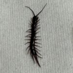 Centipede – MJAR
 Fruitfly – MJAR
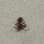 Ant – MJAR
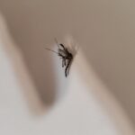 Mosquito – MJAR
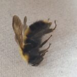 Bumblebee – MJAR