Sample information |
|
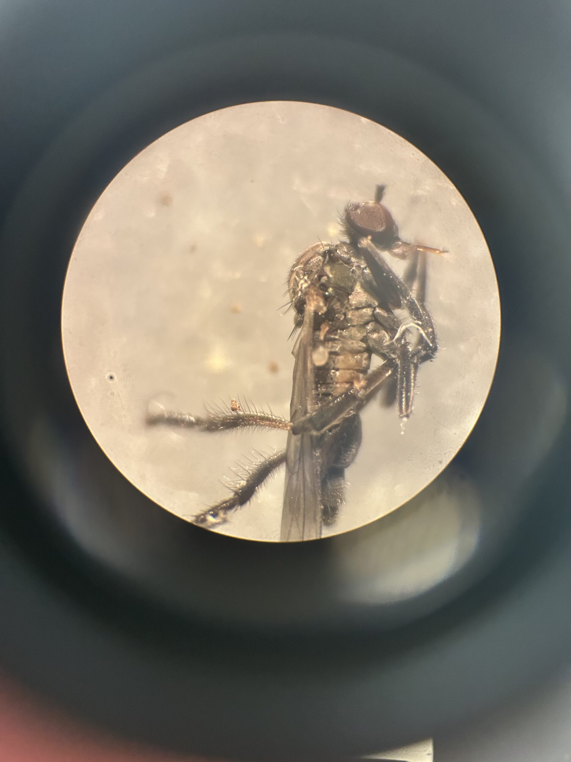
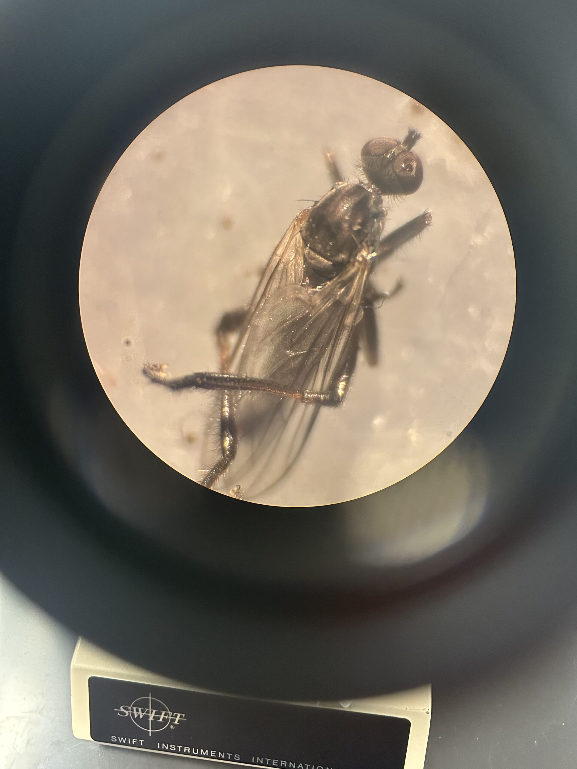
| Picture |
|
|---|---|
| Location | |
| Collection date | 04/15/2026 |
| Captive / Cultivated? | Wild-caught |
| Group | Thomas Jefferson High School for Science and Technology |
| Observations |
|
| Putative identification | Arthropoda Insecta Diptera Empididae |
Methods |
|
| Extraction kit | DNeasy (Qiagen) blood and tissue kit |
| DNA extraction location | Abdomen |
| Single or Duplex PCR | Single Reaction |
| Gel electrophoresis system | MiniPCR |
| Buffer | TBE |
| DNA stain | SYBR Safe |
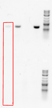
| Gel images |
|
| Protocol notes | From the Blood and Tissue Extraction protocol: Perform all centrifugation steps at room temperature (15–25°C). Redissolve any precipitates in Buffer AL and Buffer ATL. 1a. Tissue: Cut tissue (≤10 mg spleen or ≤25 mg other tissue) into small pieces, and 1c. Nucleated blood: Pipet 20 μl proteinase K into a 1.5 ml or 2 ml microcentrifuge |
Results |
|
| Wolbachia presence | No |
| Confidence level | Medium |
| Explanation of confidence level | My confidence in this result is medium, as the positive and negative controls did work. However, the top Gel band was very faint, possibly indicating not enough of the DNA sample was entered into the gel. When grinding the specimen, the exoskeleton was not ground well, possibly causing no DNA to be transmitted. |
| Wolbachia 16S sequence | |
| Arthropod COI sequence |
|
| Summary | The Empididae was found to be negative for Wolbachia. |
 Centipede – MJAR
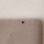 Fruitfly – MJAR
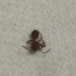 Ant – MJAR
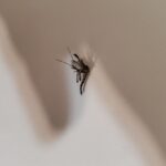 Mosquito – MJAR
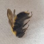 Bumblebee – MJAR