Sample information |
|||||||||||||||||
| Picture |
|
||||||||||||||||
|---|---|---|---|---|---|---|---|---|---|---|---|---|---|---|---|---|---|
| Location | Approximate location: 40.81, -77.86 |
||||||||||||||||
| Collection date | 09/28/2025 | ||||||||||||||||
| Captive / Cultivated? | Wild-caught | ||||||||||||||||
| Group | Penn State University | ||||||||||||||||
| Observations |
|
||||||||||||||||
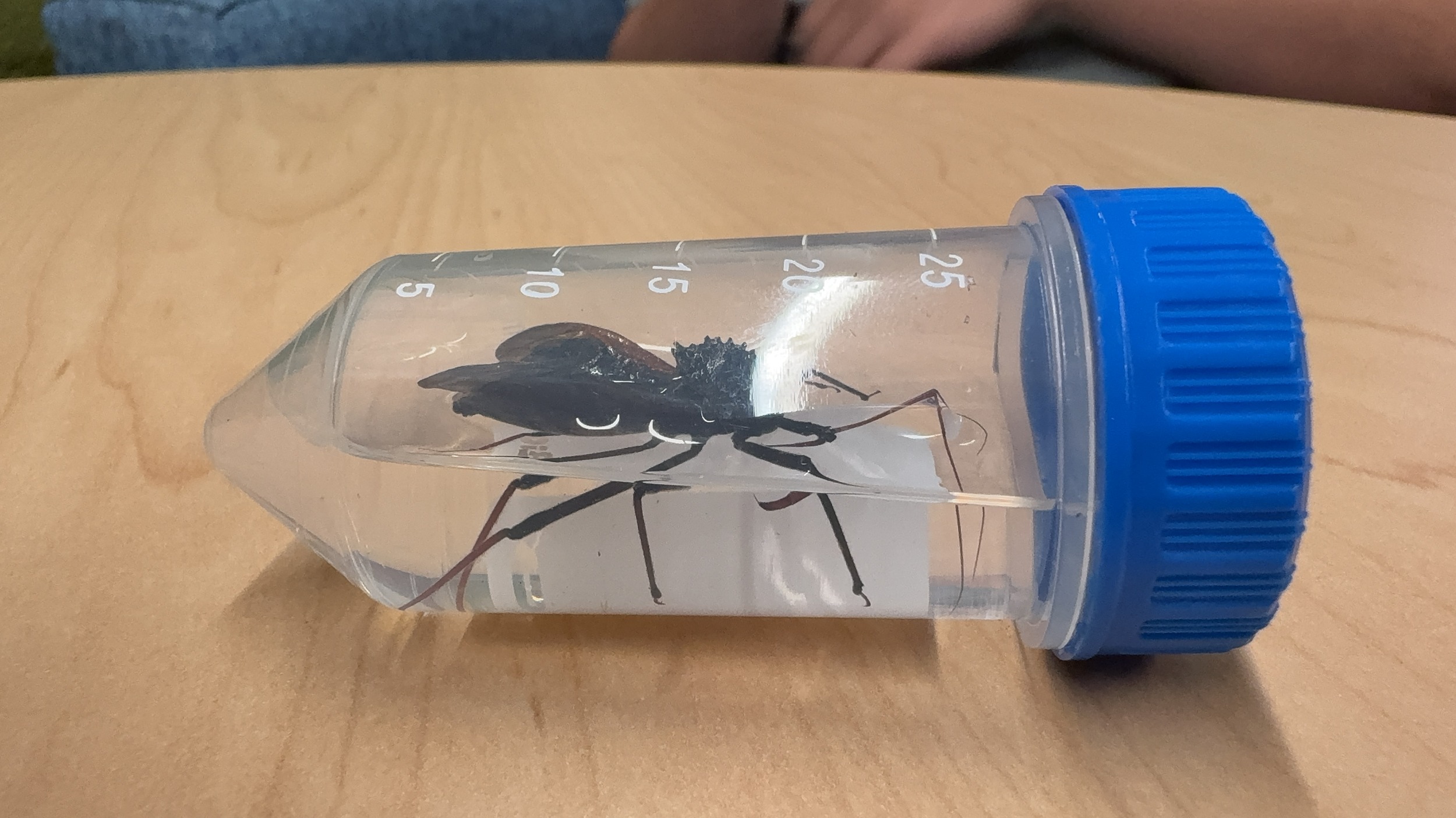
| Putative identification | Arthropoda Insecta Hemiptera Reduviidae Arilus Arilus cristatus | ||||||||||||||||
Methods |
|||||||||||||||||
| Extraction kit | QIAGEN DNeasy kit | ||||||||||||||||
| DNA extraction location | Abdomen | ||||||||||||||||
| Single or Duplex PCR | Single Reaction | ||||||||||||||||
| Gel electrophoresis system | agarose gel electrophoresis | ||||||||||||||||
| Buffer | TAE | ||||||||||||||||
| DNA stain | Ethidium Bromide | ||||||||||||||||
| Gel images |
|
||||||||||||||||
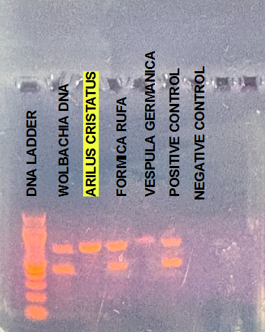
| Protocol notes | Primers The primers CO1F and CO1R were used as the arthropod-specific primers, whose main purpose was to confirm the sample tested was indeed an arthropod. Primers WSpecF and WSpecR were used as the Wolbachia-specific primers, which bound to regions of DNA that are unique to the bacterium Wolbachia Pipientis to test for its presence in the host organism. DNA extraction The arthropod was homogenized in 180 μL of ATL buffer, after which 20 μL of Proteinase K and 200 μL of AL buffer were added. The lysate was spun and incubated for 15 minutes in a 56°C water bath, after which the supernatant was moved and mixed with 200 μL of ethanol. The supernatant was then moved to a spin column, where it was washed in 500 μL of AW1 buffer and 500 μL of AW2 buffer before finally being eluted in 100 μL of AE buffer. PCR and Gel Electrophoresis 4 μL of the DNA extracted from the arthropod was mixed with 140 μL of Taq Master Mix 2x, 28 μL of PCR Water, and 21 μL each of the following primers: CO1F, CO1R, WSpecF, and WSpecR. The PCR Mix was then placed into the thermal cycler for 2 hours. The resulting PCR product was visualized by gel electrophoresis in a 1% agarose gel stained with ethidium bromide. DNA Sequencing PCR products that showed visible bands during gel electrophoresis were selected for DNA sequencing to confirm the presence of Wolbachia Pipientis. The samples were submitted for Sanger Sequencing, which is a DNA technique that confirms the nucleotide sequence of the DNA segment. This data was analyzed using BLAST (Basic Local Alignment Search Tool) through the MEGA12 (Molecular Evolutionary Genetic Analysis) database. This application constructed phylogenetic analysis for the arthropod specimens collected and compared the results to abundant arthropod species in the local area. |
||||||||||||||||
Results |
|||||||||||||||||
| Wolbachia presence | No | ||||||||||||||||
| Confidence level | High | ||||||||||||||||
| Explanation of confidence level | I am very confident that Wolbachia Pipientis was not present in my organism. The gel electropheresis we conducted had positve and negative controls to help strengthen the confidence of the results. There are bands in the positive control confirming that the PCR and gel processes were completed correctly to identify the presence of Wolbachia Pipientis. The negative control well had no bands, eliminating any suspicion of contamination. |
||||||||||||||||
| Wolbachia 16S sequence | Download AB1
|
||||||||||||||||
| Arthropod COI sequence | Download AB1
|
||||||||||||||||
| Summary | The Arilus cristatus was found to be negative for Wolbachia. | ||||||||||||||||
 (no title)
 Yellowjacket Wasp
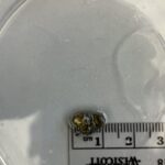 Pillbug
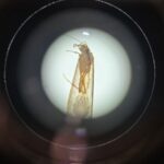 Umpachene Falls Caddisfly
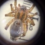 Wolf Spider