Sample information |
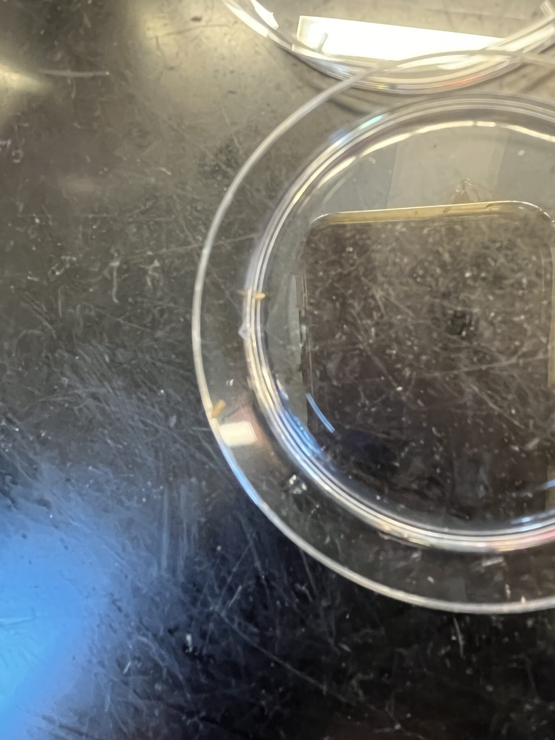
| Picture |
Entry by: JC |
| Location |
No map location data available. |
| Collection date |
10/06/2025 |
| Captive / Cultivated? |
Wild-caught
|
| Group |
Wolcott College Prep
|
| Observations |
long, thin, beige
|
| Putative identification |
Arthropoda
|
Methods |
| Extraction kit |
DNeasy (Qiagen) blood and tissue kit
|
| DNA extraction location |
Whole arthropod
|
| Single or Duplex PCR |
Single Reaction
|
| Gel electrophoresis system |
MiniPCR |
| Buffer |
TBE |
| DNA stain |
SeeGreen |
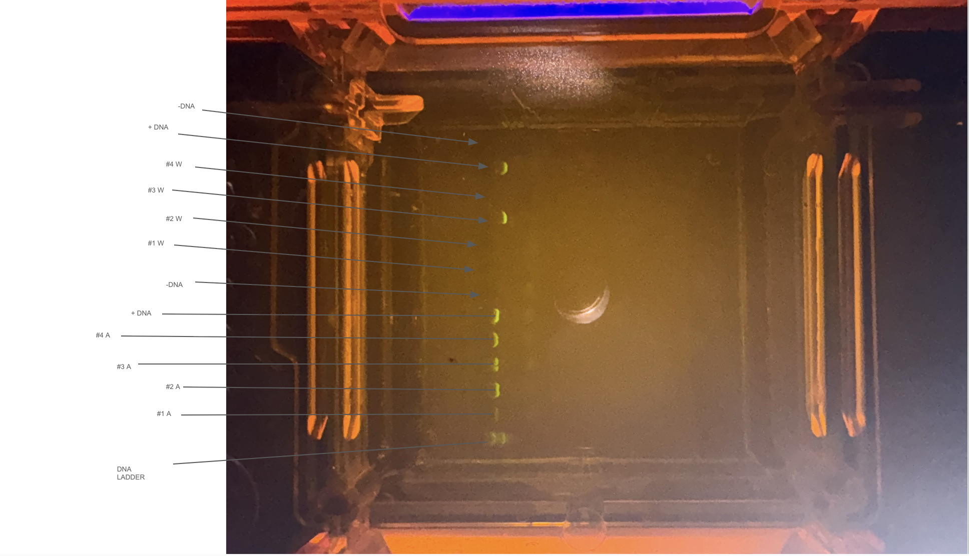
| Gel images |
|
| Protocol notes |
- Place the gel tray containing your gel in the buffer chamber
- Ensure that the clear buffer chamber is inside the blueGel electrophoresis system.
- The wells of the gel should be on the same side as the negative electrode, away from the power button.
- Use a graduated cylinder to add 30 ml of 1X TBE electrophoresis buffer
- The buffer should just cover the gel and wells.
- Ensure that there are no air bubbles in the wells (shake the gel gently if bubbles need to be dislodged).
- Starting with the ladder in lane 1, gently hover your pipette tip over the well, and load 5 uL. Follow the loading key in the table below to load the rest of your gel. Remember to change tips between each sample.
| Lane |
Sample |
| 1 |
DNA Ladder |
| 2 |
Arthropod Sample #1 |
| 3 |
Arthropod Sample #2 |
| 4 |
Arthropod (+) Control (A #3) |
| 5 |
Arthropod (-) Control (A #4) |
| 6 |
(+) DNA Control (A #5) |
| 7 |
(-) DNA Control (A #6) |
| 8 |
Wolbachia Sample #1 |
| 9 |
Wolbachia Sample #2 |
| 10 |
Wolbachia (+) Control (W #3) |
| 11 |
Wolbachia (-) Control (W #4) |
| 12 |
(+) DNA Control (W #5) |
| 13 |
(-) DNA Control (W #6) |
- Place the orange cover on the blueGel electrophoresis system
- Match the positive and negative electrode signs on the orange lid with the corresponding positive and negative signs on the blue base.
- The orange lid should sit flush with the blue base using little force.
- Press the “Run” button
- Check that the green light beside the power button remains illuminated.
- Conduct electrophoresis for 20-25 minutes
- The colored dye should progress to about half the length of the gel.
- Longer electrophoresis times will result in better size resolution.
- Press the “light bulb” button to turn on the blueGel transilluminator.
- For best viewing, place the viewing chamber over the blueGel machine.
- Gels may be viewed at the end of the run or periodically throughout the run.
- Ensure that the bands in your gel have separated enough to clearly interpret your results
- Run the gel longer if needed to increase resolution.
- Take a picture and insert it in the correct post-lab question.
|
Results |
| Wolbachia presence |
No
|
| Confidence level |
Medium
|
| Explanation of confidence level |
Some of the controls were not what they were supposed to be however some are correct.
|
| Wolbachia 16S sequence |
|
| Arthropod COI sequence |
|
| Summary |
The Arthropoda was found to be negative for Wolbachia.
|
 (no title)
 Yellowjacket Wasp
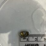 Pillbug
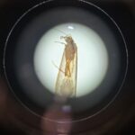 Umpachene Falls Caddisfly
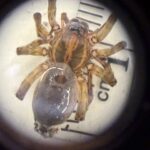 Wolf Spider