Sample information |
|||||||||||||||||||||||||||||
| Picture |

|
||||||||||||||||||||||||||||
|---|---|---|---|---|---|---|---|---|---|---|---|---|---|---|---|---|---|---|---|---|---|---|---|---|---|---|---|---|---|
| Location | |||||||||||||||||||||||||||||
| Collection date | 10/06/2025 | ||||||||||||||||||||||||||||
| Captive / Cultivated? | Wild-caught | ||||||||||||||||||||||||||||
| Group | Wolcott College Prep | ||||||||||||||||||||||||||||
| Observations | We took a standard size fly and removed the inside of the abdomen. We are unsure whether wolbachia is present or not. |
||||||||||||||||||||||||||||
| Putative identification | Arthropoda Insecta Diptera Muscidae Musca Musca domestica | ||||||||||||||||||||||||||||
Methods |
|||||||||||||||||||||||||||||
| Extraction kit | DNeasy (Qiagen) blood and tissue kit | ||||||||||||||||||||||||||||
| DNA extraction location | Abdomen | ||||||||||||||||||||||||||||
| Single or Duplex PCR | Single Reaction | ||||||||||||||||||||||||||||
| Gel electrophoresis system | MiniPCR | ||||||||||||||||||||||||||||
| Buffer | TBE | ||||||||||||||||||||||||||||
| DNA stain | SeeGreen | ||||||||||||||||||||||||||||
| Gel images |

|
||||||||||||||||||||||||||||
| Protocol notes |
|
||||||||||||||||||||||||||||
Results |
|||||||||||||||||||||||||||||
| Wolbachia presence | Unknown | ||||||||||||||||||||||||||||
| Confidence level | Low | ||||||||||||||||||||||||||||
| Explanation of confidence level | Since the A2 control arthropod does not show up with a line, it is unknown whether wolbachia was present in the abdomen or not. The wolbachia column (W2) is blank but we are unsure if this is because there is no wolbachia or if there was a mistake with the procedure. |
||||||||||||||||||||||||||||
| Wolbachia 16S sequence | |
||||||||||||||||||||||||||||
| Arthropod COI sequence |
|
||||||||||||||||||||||||||||
| Summary | |||||||||||||||||||||||||||||
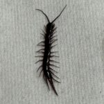 Centipede – MJAR
 Fruitfly – MJAR
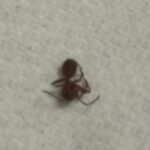 Ant – MJAR
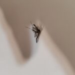 Mosquito – MJAR
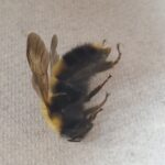 Bumblebee – MJAR