Sample information |
|
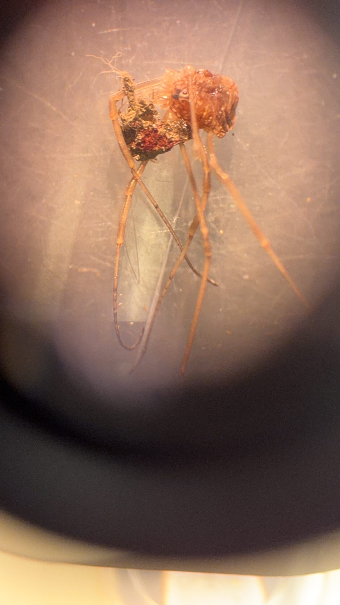
| Picture |
|
|---|---|
| Location | |
| Collection date | 04/15/2026 |
| Captive / Cultivated? | Wild-caught |
| Group | Thomas Jefferson High School for Science and Technology |
| Observations |
|
| Putative identification | Arthropoda Insecta Diptera Rhagionidae Chrysopilus Chrysopilus basilaris |
Methods |
|
| Extraction kit | DNeasy (Qiagen) blood and tissue kit |
| DNA extraction location | Whole arthropod |
| Single or Duplex PCR | Single Reaction |
| Gel electrophoresis system | MiniPCR |
| Buffer | 1X TBE |
| DNA stain | SYBR Safe |
| Gel images |
|
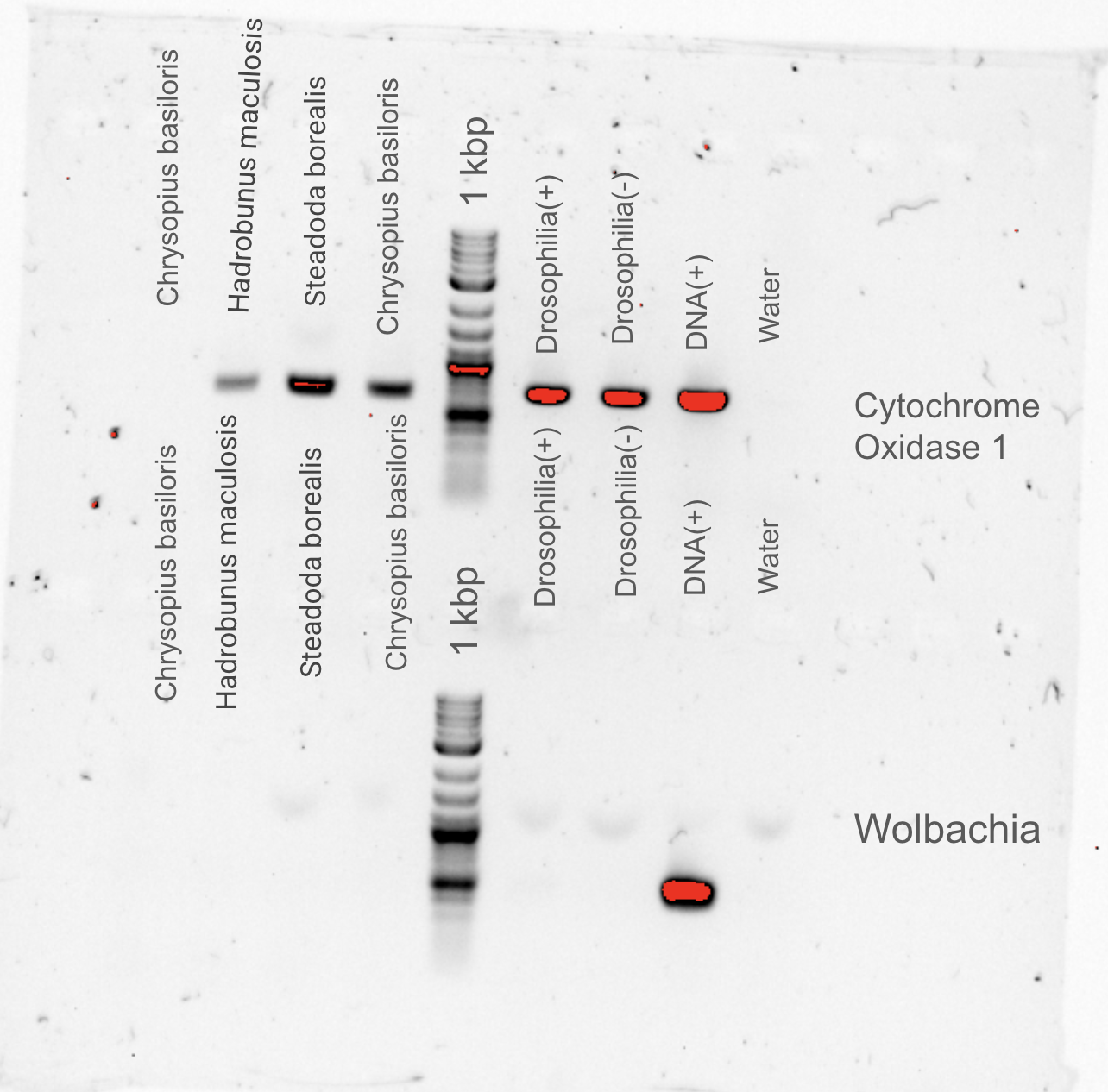
| Protocol notes | DNA Extraction: I made sure to crush the arthropod really well and diluted my sample to meet the requirements for PCR. We ran gel electrophoresis for both Cytochrome c Oxidase subunit from Drosophila (top of the gel) and for Wolbachia-specific DNA from Drosophila (bottom of the gel) for four arthropod samples. Chrysopius basiloris results are in the 4th lane for top and bottom gels labeled Chrysopius basiloris. |
Results |
|
| Wolbachia presence | No |
| Confidence level | High |
| Explanation of confidence level | All of my controls worked as expected, and I had no problems with the protocols. I wasn’t surprised by any of the bands on the gel; therefore, I am confident that the snipe fly wasn’t infected with Wolbachia. |
| Wolbachia 16S sequence | |
| Arthropod COI sequence |
|
| Summary | The Chrysopilus basilaris was found to be negative for Wolbachia. |
 We found this near a grassy area with a moderate density of trees. I collected it in the morning around 11 AM, and the weather was sunny and around 85º F. There were many snipe flies in the grassy area, but I only collected 1.
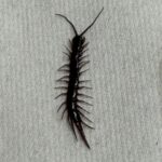 Centipede – MJAR
 Fruitfly – MJAR
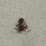 Ant – MJAR
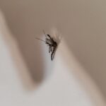 Mosquito – MJAR
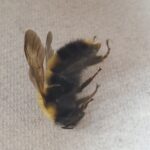 Bumblebee – MJAR