Sample information |
|
| Picture |
|
|---|---|
| Location | |
| Collection date | 11/13/2024 |
| Captive / Cultivated? | Wild-caught |
| Group | Hawaii Community College |
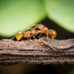
| Observations | Fire ant was captured using peanut butter in a vile. A small quantity of peanut butter; no more than pea-sized was placed in a vile and left for an hour. Over 20 specimens were collected. This site gave me goosebumps. |
| Putative identification | Arthropoda Insecta Hymenoptera Formicidae Wasmannia Wasmannia auropunctata |
Methods |
|
| Extraction kit | tweezers |
| DNA extraction location | Whole arthropod |
| Single or Duplex PCR | Single Reaction |
| Gel electrophoresis system | Standard electrophoresis system |
| Buffer | TBE |
| DNA stain | SYBR Safe |
| Gel images |
|
| Protocol notes | The extraction methods include washing the specimen, drying it, then using a pestle and lysis buffer to break it up. Then I heated the tubes, cooled them, centrifuged it then separated the debris and transferred the supernatant to a new tube. The supernatant was then combined with isopropanol, mixed, and centrifuged to get a pellet. The pellet was then washed with ethanol, and then air dried. The DNA pellet was then resuspended in the elution buffer and incubated at 65 degrees C. It was then stored at -20 degrees C. To set up the PCR we would have put on PPE and cleaned the work station. We then would have labeled the DNA we extracted as well as the PCR tubes with t=our initials and sample numbers.
For the CO1 gene we would have used a mixture containing taq polymerase, CO1 primers, dNTPs, and buffers. Then adding 2 ul of DNA into the mixtures in the labeled PCR tubes. The lids were then secures, centrifuged, then placed in the PCR rack. The PCR cycle for CO1 consists of an initial denaturation for 2 minutes at 94 degrees C, followed by 30 cycles of 30 seconds at 94 degrees, annealing for 45 seconds at 49 degrees C and then extension for 60 seconds at 72 degrees C.Then it was extended for 10 minutes at 72 degrees for the final extension.
For the Wolbachia sample we would have used PCR mixture containing taq polymerase, Wspec primers, dNTPs, and buffers. We would have added 2 uL of DNA sample into the labeled PCR tubes, centrifuged them, then placed them on the PCR rack. The PCR cycle would have then been performed the same with the exception of the annealing stage which was 45 seconds at 55 degrees Celcius. The samples were then stored in the freezer and the workstations were cleaned. As always this began by donning PPE and cleaning the work stations. The casting tray was then prepared by putting the rubber caps on each end and inserting the combs. The agarose solution was then prepared by mixing 1 gram of agarose powder, 50 mL of 1X TBE buffer, and dissolved by heating and swirling the mixture to make a 2% gel to create the optimal sized pores for this electrophoresis test. The gel was then cooled before adding 2.5 uL of SYBR Safe DNA stain into the gel mixture. We then poured the mixture into the casting block and allowed it to cool. The electrophoresis was then set up by removing the gel block and placing it into the electrophoresis chamber and submerging it with 300 mL of TBE buffer. We then created a loading key table listing the lanes and the sample IDs. The gel was then ready to be loaded and ran. To run the agarose gel we first practiced loading. This step is optional. We then prepared the sample by placing a piece of parafilm on the bench and labeling it with the sample IDs. We then added 2 uL of 6x DNA loading dye to the designated parafilm and mixed 10 uL of DNA ladder or PCR samples with the loading dye. The DNA ladder and samples were then loaded into the wells ensuring to keep track of which sample went in each well/lane.
Following this, we ran the gel by connecting the negative and positive electrodes to the power supply. We then ran the gel at 130 volts for 30 minutes.
After the electrophoresis was completed, we turned off the power and disconnected the electrophoresis device. The gel was then placed on a UV light transilluminator and analyzed using eye protection to observe the results of the test. The results were then recorded and the stations were cleaned up. |
Results |
|
| Wolbachia presence | Yes |
| Confidence level | Medium |
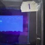
| Explanation of confidence level | My confidence level is medium because my results showed 1/2 of the samples were positive for the CO1 gene but 2/2 were positive for the 16S rRNA gene. Lane 1: 1kb DNA Ladder. Lane 2: CO1 A (sample 1) Band present: No Lane 3: CO1 B (Sample 2) Lane 4: Wolbachia A (Sample 1) Lane 5: Wolbachia B (Sample 2) |
| Wolbachia 16S sequence | Download AB1
|
| Arthropod COI sequence | Download AB1
|
| Summary | The Wasmannia auropunctata was found to be postive for Wolbachia. |
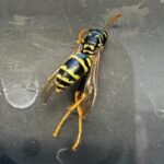 European Wasp
 Small Honey Ant
 7A
7A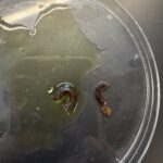 Millipede 7F