Sample information |
|
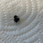
| Picture |
|
|---|---|
| Location | |
| Collection date | 10/11/2024 |
| Captive / Cultivated? | Wild-caught |
| Group | Owego Free Academy |
| Observations | |
| Putative identification | Arthropoda |
Methods |
|
| Extraction kit | DNeasy (Qiagen) |
| DNA extraction location | Abdomen |
| Single or Duplex PCR | Single Reaction |
| Gel electrophoresis system | MiniOne |
| Buffer | TBE |
| DNA stain | GelGreen |
| Gel images |
|
| Protocol notes | |
Results |
|
| Wolbachia presence | Yes |
| Confidence level | Medium |
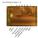
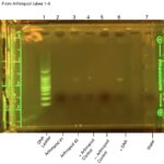
| Explanation of confidence level | In the Arthropod DNA gel, we were not able to extract DNA of Arthropod #1, we didn’t vortex the PCR tube, so I believe that we didn’t pick up the DNA with the micropipette. Switched Wolbachia positive control with Wolbachia negative control in the Wolbachia gel electrophoresis. Summary: In figure two: the gel electrophoresis results from the arthropod DNA is shown. In lane 1 the DNA ladder is shown with bright bands. This means that the Gel electrophoresis machine was working. In lane 2 and 3, it shows both of the arthropod DNA that we collected. However we were only able to extract arthropod DNA from arthropod #2. We know this because the gel is only highlighted in lane 3 and not lane 2. We believe that lane 2 didn’t have a band because we didn’t vortex the pcr tube enough, so the micropipette didn’t pick up the DNA. In lanes 4 and 5, we have the arthropod positive and negative controls. In the negative control lane we don’t see a band and in the positive lane we do see a band. This is because the negative control didn’t have arthropod DNA and the positive control does have arthropod DNA. We use these controls to make sure that our data is valid. In lanes 6 and 7, we see the DNA positive control and the water. We see a band in the positive DNA control and no arthropod DNA in the water control. This is because the DNA control was a positive control and there was no DNA in the water, which also means there is no contamination. In figure three: We see the results of the Wolbachia gel electrophoresis. In lane 1 we see the DNA ladder, which is there to make sure that the gel electrophoresis machine is working. In lanes 2 and 3 that they both have bands; which shows that Wolbachia is present in our arthropods. This is what leads us to believe that we didn’t vortex the pcr tube with arthropod #1 DNA (in the first gel testing for Arthropod DNA) instead of not macerating it enough; because the band in the arthropod #1 lane shows that there was DNA extracted from the pcr tube. This matches up with the results from figure two where we only saw DNA from arthropod two. In lanes 4 and 5 we see the two controls for the arthropod DNA. We see that the negative control has a band and the positive control doesn’t have a band. This leads us to believe that we switched the tubes with each other when putting the DNA into the gel electrophoresis machine. In lanes 6 and 7, we see that there is a band where the positive DNA control is and no band where the water is. This is because the water was a negative control that had nothing in it, and the dna control was guaranteed to have Wolbachia DNA in it. These controls are there to validate our data and make sure there was no contamination. |
| Wolbachia 16S sequence | |
| Arthropod COI sequence |
|
| Summary | The Arthropoda was found to be postive for Wolbachia. |
 Wolbachia Project
 Alexander Devlin-Myrmica
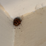 Brookelynn Foote- Coccinellidae
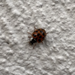 Maile Bentz- Harmonia axyridis
 Madison Adams – Harmonia axyridis