Sample information |
|
| Picture |
|
|---|---|
| Location | |
| Collection date | |
| Captive / Cultivated? | Wild-caught |
| Group | California Academy of Mathematics and Science |
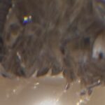
| Observations | Approx 5mm x 3mm x 2.5mm, exoskeleton consisting of overlapping blue-gray shells with white-green longitudinal spot pattern |
| Putative identification | Arthropoda Malacostraca Isopoda |
Methods |
|
| Extraction kit | DNeasy (Qiagen) blood and tissue kit |
| DNA extraction location | Gonads dissected |
| Single or Duplex PCR | Single Reaction |
| Gel electrophoresis system | Standard electrophoresis system |
| Buffer | TAE |
| DNA stain | UView |
| Gel images |
|
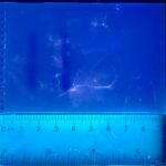
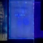
| Protocol notes | Run gels at 100V constant for 45 minutes using HindIII ladder mixed with UView. |
Results |
|
| Wolbachia presence | No |
| Confidence level | High |
| Explanation of confidence level | The arthropod DNA sequences (CO1) showed up for the insect and DNA positive samples, which not showing for the DNA negative control. This indicates that DNA extraction, PCR, and gel electrophoresis were correctly conducted without contamination, validating the procedure used for the Wolbachia 16S sequence electrophoresis. The results are further justified by the DNA positive and Wolbachia positive samples being the only samples to show a band indicating the 16S sequence on the Wolbachia PCR gel, meaning that there was no contamination specifically during the DNA extraction, PCR, and gel electrophoresis protocol concerning the Wolbachia 16S sequence. |
| Wolbachia 16S sequence | |
| Arthropod COI sequence |
|
| Summary | The Isopoda was found to be negative for Wolbachia. |
 EG Wolbachia 7L
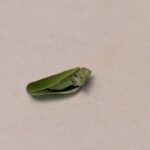 7A
7A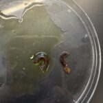 Millipede 7F
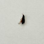 Coleoptera
 Emily Yocum- Reticulitermes flavipes