Sample information |
|
| Picture |
|
|---|---|
| Location | |
| Collection date | 11/21/2024 |
| Captive / Cultivated? | Wild-caught |
| Group | Hawaii Community College |
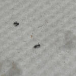
| Observations | Small black ants provided by instructor; must have been easy to catch. |
| Putative identification | Arthropoda |
Methods |
|
| Extraction kit | pcr |
| DNA extraction location | Whole arthropod |
| Single or Duplex PCR | Single Reaction |
| Gel electrophoresis system | Standard electrophoresis system |
| Buffer | TBE |
| DNA stain | SYBR Safe |
| Gel images |
|
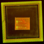
| Protocol notes | Gel Lanes (Top)
Experimental Set up The collection of two insect specimens were subjected to DNA extraction for analysis under the process of cell lysis, DNA precipitation, and DNA elution. Specimens were rinsed with water to remove excess ethanol, and then transferred into two separate 1.5 mL microcentrifuge tubes. A sterile pestle grinds the sample after a 200 µL Cell Lysis Buffer was added, ensuring optimal maceration. To rid of debris, the samples were heated to 95°C and incubated approx. 5 minutes and centrifuged for 2 minutes. The results contained a supernatant that is then precipitated using cold isopropanol (150 µL) and centrifuged for 5 minutes. Products are air-dried and rid of supernatants whilst ensuring that the DNA pellets are left behind. Pellets are then washed with 100 µL of 70% ethanol. 50 µL of DNA Elution Buffers are added to each tube and incubated in a 65°C water bath for approx. 5 minutes. Once incubated, 2 µL of the DNA extracted are extracted to pre-aliquoted PCR mixes for the appropriate arthropod CO1 and wolbachia 16S rRNA gene. The PCR tubes are then amplified in a thermal cycler using appropriate steps with optimal temperature DNA amplification. To prepare for visualizing the extraction of the potential DNA and bacteria, a 2% agarose gel would be created by dissolving 0.5g of agarose powder in 25mL of a 1XTBE buffer, cooled, and then poured into a tray. Samples would be mixed with heavy loading die, loaded into the vents, and would undergo electrophoresis on a 2% agarose gel mixed with SYBR Safe DNA dye. The test would be run at 130V for ~21 minutes. Bands were visualized in a transilluminator and photographed for analysis. |
Results |
|
| Wolbachia presence | Yes |
| Confidence level | High |
| Explanation of confidence level | The controls showed great results. The presence of DNA in the arthropod CO1 samples displayed the correct band size, indicating a successful extraction. The presence of wolbachia is seen with the correct band size, indicating that the insects provided were positive for wolbachia. |
| Wolbachia 16S sequence | Download AB1
|
| Arthropod COI sequence | Download AB1
|
| Summary | The Arthropoda was found to be postive for Wolbachia. |
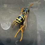 European Wasp
 Small Honey Ant
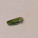 7A
7A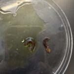 Millipede 7F