Sample information |
|
| Picture |

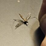
|
|---|---|
| Location | |
| Collection date | 11/13/2024 |
| Captive / Cultivated? | Wild-caught |
| Group | Hawaii Community College |
| Observations | During the experiment after smashing the insect with the sterile pestle and adding the sample into the centrifuge there was still a giant pellet at the bottom of the test tube. I returned the sample to the centrifuge and the giant pellet still remained. I Pipetted out as much as I could without disturbing the pellet and even after the third round in the centrifuge and other experimental steps the pellet still remained. |
| Putative identification | Arthropoda Insecta Diptera Tipulidae |
Methods |
|
| Extraction kit | pcr |
| DNA extraction location | body without wings |
| Single or Duplex PCR | Single Reaction |
| Gel electrophoresis system | Standard electrophoresis system |
| Buffer | TBE |
| DNA stain | SYBR Safe |
| Gel images |

|
| Protocol notes | Summary of events: During the experiment I was briefly educated by my classmates that the bug I had was NOT a mosquito but a cane fly. That did not change the fact that I did have to dissect the bug and tear apart its little legs and wings using tweezers. After the budget dissection I added the bug torso to a test tube and added, cell lysis buffer. Using a sterile pestle I crushed the bug in the test tube making sure to push it hard against the bottom twisting as I pushed, then I dragged it up against the side of the test tube making sure it was thoroughly squished but parts of the bug did get stuck on the pestle and I did have to rub it off on the rim of the test tube to get it off. In the process the cell lysis buffer bubbled and disappeared a little while the otherwise clear lysis buffer coloring was brown and murky. Added the test tube to the cooling block, set my phone timer for 5 minutes and waited. After 5 minutes I collected my test tube and tilted my test tubes back and forth for a few minutes. I added them to the mini centrifuge for 2 minutes and began preparing my isopropanol test tube by pipetting and marking the test tube. After the two minutes finished I started to pipette the supernatant out of the test tube and into another test tube. There were some issues where some of the bug parts dislodged from the bottom of the test tube and I needed to put it back in the centrifuge for another 2 minutes. After that there were no issues collecting the supernatant. From there I added the supernatant to the isopropanol and mixed them together by once again flipping the test tube a few times. After that I placed the newly mixed tube into the centrifuge once again but this time for 5 minutes. After the time was up I collected my test tube and noticed that there was a large visible pellet at the bottom of the tube. It did not move and wouldn’t dislodge in the final steps. I pipetted the liquid out of the test tube and into a small waste beaker and balanced the test tube upside down on a paper towel for a few minutes. Afterwards, I added more isopropanol to the test tube, flipped it back and forth some more to mix it once again and put it back into the centrifuge for a minute. After a minute I removed the test tubes from the centrifuge and added some DNA elution buffer, labeled my test tubes and handed it off to the instructor. |
Results |
|
| Wolbachia presence | No |
| Confidence level | Medium |
| Explanation of confidence level | This is my first time semester taking a lab class and it is entirely possible that I made mistakes during this experiment so I cannot say for certain that my results are as they should be. |
| Wolbachia 16S sequence | |
| Arthropod COI sequence |
|
| Summary | The Tipulidae was found to be negative for Wolbachia. |
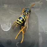 European Wasp
 Small Honey Ant
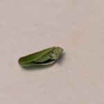 7A
7A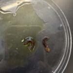 Millipede 7F