Sample information |
|
| Picture |
|
|---|---|
| Location | |
| Collection date | 09/06/2024 |
| Captive / Cultivated? | Wild-caught |
| Group | Owego Free Academy |
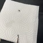
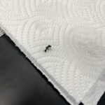
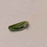
| Observations | The Ants looked like normal ants, they were found in a grassy area, with some rocks. |
| Putative identification | Arthropoda Insecta Hymenoptera |
Methods |
|
| Extraction kit | DNeasy (Qiagen) |
| DNA extraction location | Rear half |
| Single or Duplex PCR | Single Reaction |
| Gel electrophoresis system | MiniOne |
| Buffer | |
| DNA stain | GelGreen |
| Gel images |
|
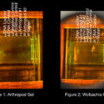
| Protocol notes | When our group had put the comb into the tray that had our melted gel in it, the comb wasn’t even within the tray. This resulted in our gel coming out with uneven wells throughout the gel. |
Results |
|
| Wolbachia presence | Yes |
| Confidence level | Medium |
| Explanation of confidence level | Confidence Level
Summary Our Arthropod shown in figure 1 shows the crushed up arthropods, and got DNA from them. While looking at the Gel you can see that the DNA moved out of the well and down the gel, however that shows there’s DNA. Because of the comb malfunction described more in the protocol notes, the gel electrophoresis wells didn’t form correctly in 1-6, leading to 7-9 only forming correctly. Even though 1-6 wells didn’t conclude any evidence, DNA was still injected into the wells. In well 1 and 2 we left empty because we only had to fill 7 wells and those two wells were the shallowest out of them all. Well 3 was our DNA ladder, and in 4 was the control DNA, neither were present after the 20 minutes. Well 5 was the negative Arthropod control, and 6 was the positive arthropod control, neither showed lines because the wells were shallow and DNA floated out. The 7th well was the Arthropod 1, and it showed a line, showing we crushed up the bug and did the PCR correctly. The 8th well contained Arthropod #2 which also showed a line, meaning we followed the PCR correctly. The last well (9th one) was water, in this case you wouldn’t want to see a line because that would mean contamination has occurred, so we did that correctly too.
Figure 2 shows a gel electrophoresis for wolbachia. In the first well is the DNA ladder, 2nd was Arthropods 1, 3rd was arthropod 2, which showed a line illustrating wolbachia present, 4th well was negative control, 5th was the positive control, meaning everything worked correctly, because we knew it came to us with wolbachia. 6th DNA control, 7th was water which didn’t have a line, meaning there wasn’t any contamination within the lab.
|
| Wolbachia 16S sequence | |
| Arthropod COI sequence |
|
| Summary | The Hymenoptera was found to be postive for Wolbachia. |
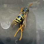 European Wasp
 Small Honey Ant
 7A
7A Millipede 7F